

Tomography is a new NDT technology (Non Destructive Testing) to obtain a 3D reconstruction of internal defects. Faults can thus be visualized and quantified with precision (spatial position, area, shape factor, ...).
What is the principle of tomography?
Tomography is to use a source radio (microwave hearth) on a workpiece or a sample rotation. The 3D image is then reconstructed by calculation.

Compared to other analysis of internal defects, the tomography means has advantages:
- True 3D image to visualize and diagnose finely internal defects,
- Quantification (Part geometry and defects)
But also imposes constraints:
- Cost of the tool
- Analysis time.
That allows to analyze the CT?
Tomography can do two things:
- From 3D dimensional control room
- From internal health control room
A founder may equip it?
Yes, some have begun to do so for dimensional inspection and health room. Customers or R & D centers are also equipped with a tomograph.
Quantification of internal defects
Micro-tomography, used by IWTC in an R & D has to acquire images with a resolution between 3 microns and 20 microns (1 voxel = 20 mm). Different materials were analyzed (Al Si9Cu3 Al Si12, Al Si17Cu3, zamak, magnesium, ductile iron, aluminum foam). The vast majority concerned the diecasting (Al, Zn) on the draft R & D.
Tomography provides access to the following information:
- Porosity rate (in an area of each)
- Pore diameter (average, min, max)
- Shape factor of pores
- Distance of the pores of the skin piece. This parameter is of crucial importance for the fatigue (in alternating bending, but also to a lesser extent in tension / compression) in the sense that defects very close to the surface initiate fatigue cracks.
However, all these data require an operation manual recount:
- Isolate the area of interest
- Perform a thresholding operation (transformation of an initial gray level image into a black-white binary image). The thresholding operation (image analysis) is clearly the most delicate.
- Erosion and dilation operations that eliminate noise (resulting from thresholding) and too small defects to have a number of pores easily analyzable.
- Analysis of the proper file for realistic rendering and transfer to an Excel spreadsheet.
Image processing
Analysis and quantification of pores
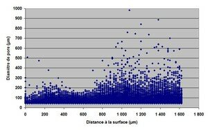
Distance to the surface (pore diameter and the ordinate) for an aluminum part 3.5 mm thick

Pores with respect to the workpiece surface (left and right) part.
Area skin part is relatively healthy.
Images of 3D tomography defects
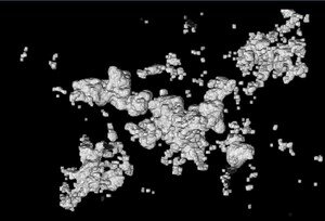
Micro-shrinkage (3D view) - Shrinkage
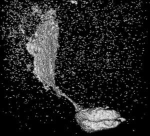
Recovery (3D view) - Cold shut
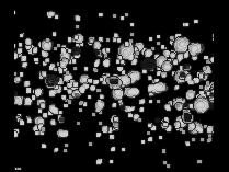
Blowhole (3D view) - Blowhole pores
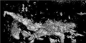
Shrinkage (3D view) without filtering - shrinkage (without erode and dilate operations)

Shrinkage (Al Si9Cu3)
Vídeos
A means of CND future
Tomography is a means of control that begins to emerge from the R & D and is led to the industrial development in the future as to access additional information relative to the fluoroscopy.
Source: My little blog fonderie


